
病理形態解析支援
班長 二口 充(山形大学)
活動内容
齧歯類(マウス・ラット)、遺伝子改変動物、ゼブラフィッシュなどの魚類・カエルをはじめとする真核生物において、腫瘍性病変をはじめ、炎症性疾患・神経変性疾患・老化など、生命体個体のすべての系統を対象とした病理形態学的解析を支援する。メンバー全員が日本病理学会認定病理専門医であり、HE染色・特殊染色・免疫染色・in situ hybridizationまで幅広く対応する。iPS細胞由来の動物モデル解析にも専門家が対応可能である。
支援の特性
ヒト疾患を熟知した病理形態学の専門家集団であることが、本支援の最大の強みである。動物モデルの所見をヒト疾患の文脈で解釈できるため、基礎研究と臨床をつなぐ視点から支援が可能である。また、従来の形態評価にとどまらず、分子データや画像解析技術との統合など、病理形態学の新たな展開にも対応しており、研究の深化と新規発見を支える。
支援内容
個体の病理形態学的解析支援
齧歯類を中心とした真核生物において、腫瘍性病変をはじめ、炎症性疾患・神経変性疾患・老化など、生命体個体のすべての系統を対象とした解析に対応する。メンバー全員が日本病理学会認定病理専門医であり、iPS細胞由来の動物モデル解析にも対応可能である。
病理形態解析のための染色支援
HE染色・特殊染色・免疫染色・in situ hybridizationまで幅広く対応する。形態解析データの定量化支援も行う。
動物モデルとヒト疾患の対応検討
薬剤投与に伴う臓器の形態学的変化を解析し、ヒト疾患との対応関係を検討する。ヒト病理を熟知した専門家が対応するため、動物モデルに対応するヒト疾患の高精度な同定が可能である。
胎児死亡・腫瘍形成能に関する解析支援
iPS細胞を含む先進的な再生医学・発生工学的手法に伴う発生異常や腫瘍形成能の解析を得意とする。奇形腫のみならず、胎児の先天性奇形や死亡原因の同定にも対応する。
環境因子・微小環境に着目した病理解析支援
アスベストやマイクロ・ナノプラスチックなど環境因子が個体に与える影響を、鉄・酸化ストレス代謝の観点から動物モデルで解析する支援を行う。トランスクリプトミクス・メタボロミクスと組織学的所見を統合した解析に対応する。
特殊染色・免疫組織化学・免疫蛍光染色による形態解析を分子・微小環境データと統合し、オミクスデータを形態学に落とし込む包括的な病理解釈支援を提供する。
デジタル病理画像の定量解析・AI画像解析支援
AI画像解析技術を用いて、従来の病理組織学的評価を定量化・客観化する支援を行う。HE染色画像のニューラルネットワークによる形態クラスタリングや、免疫染色における目的細胞集団の定量化などに対応する。
「見たいものしか見えない」評価から「見えなかったものを可視化する」評価へ--客観的手法を加えることで、新規知見の発見につながる解析支援を提供する。
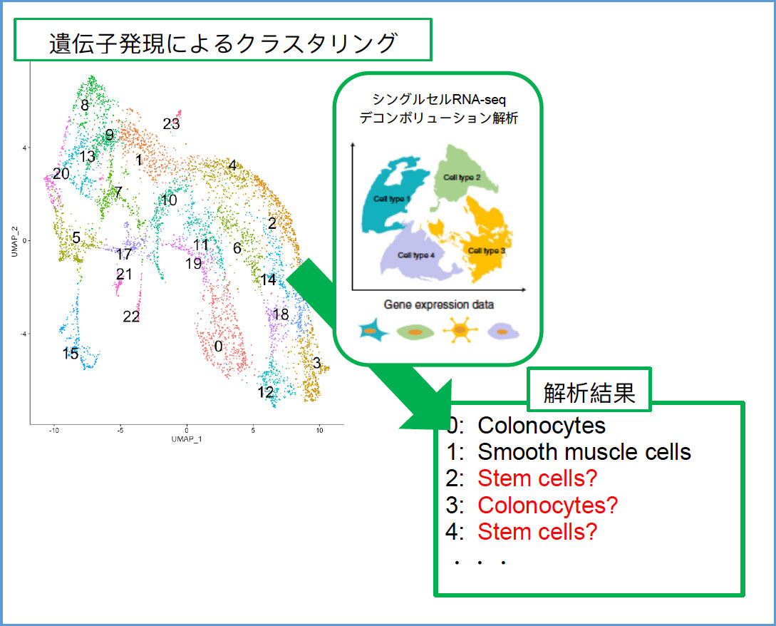
空間トランスクリプトーム解析における組織像同定支援
空間トランスクリプトーム解析において、遺伝子発現パターンによるクラスター分類の構成細胞同定を病理組織学的観点から支援する。遺伝子発現解析では同定困難なクラスターに対しても構成細胞の情報を提供し、解析の広がりに貢献する。
※空間トランスクリプトーム解析におけるクラスター解析までの行程は、支援対象ではありません。
注目の支援 酸化ストレス・鉄代謝・フェロトーシスに着目した病理解析
注目の支援 神経疾患モデルマウスの脊髄病変の病理形態解析
注目の支援 iPS細胞の使用に関連した病変の解析(腫瘍病変・分化など)
注目の支援 肝腫瘍などのホルマリン固定パラフィン包埋(FFPE)標本からの核酸・タンパク質の抽出・解析ならびに電顕による解析
以下のようなご相談を個別にメールで受け付けております。事務局までご連絡ください。
- 遺伝子改変動物に肉眼的な変化があるが、どのような病変かわからない。
- 動物個体の死亡原因を知りたい。
- 動物モデルのヒトにおける対応疾患を知りたい。
- 動物モデルにおいて、すべての臓器に形態学的な変化がないことを確認したい。
- 遺伝子改変動物が胎児期に死亡するが、その原因を突き止めたい。
- 免疫染色がうまくいかない。染色と結果の評価を依頼したい。
- プレパラートの画像をデジタル化し、virtual slideを作製したい。
- WSI(whole slide imaging)を用いたAI画像解析を行いたい。
など、種々のご要望にお応えします。まずはご相談ください。
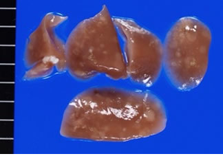 Starting from macroscopic findings |
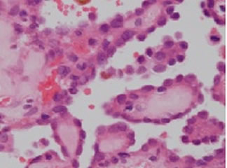 Histology with accurate diagnosis |
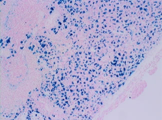 Important information with special staining |
 Analysis of embryo with serial sections |
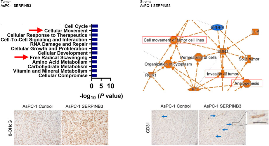 オミクス解析で示唆された酸化ストレスや血管新生の変化を免疫染色(8-OHdG、CD31)で可視化。 |
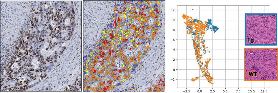 デジタル化病理画像の定量や人工知能を活用した画像解析 |
 病理組織学的に細胞・組織特定を支援します |
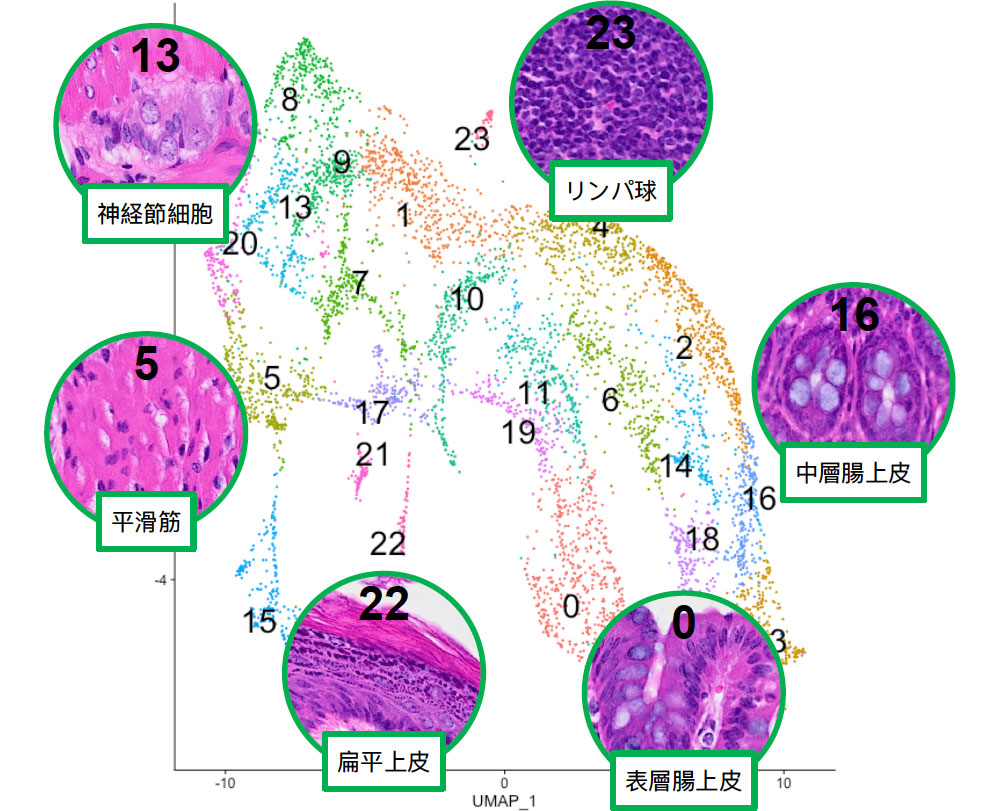 各クラスターの領域に基づいた典型的組織像の特定 |
担当者一覧 Members for this support
二口 充(山形大学)
神田 浩明(埼玉県立がんセンター)
宮崎 龍彦(岐阜大学医学部附属病院)
高松 学(がん研究会・がん研究所)
鈴木 周五(大阪公立大学)
荒木亜寿香(山口大学)
大原 悠紀(名古屋大学)
支援申込みフォーム