


What is congenital cerebral hypomyelination?
Congenital cerebral hypomyelination is the common name for a group of brain disorders in children, which occur due to developmental defects in the white matter of the brain (described in more detail below). At present, eleven such diseases are known (see diagnostic criteria). The symptoms vary depending on the patient's condition. In most cases, developmental delay, abnormal flicking of the eyes (known as nystagmus), and gradual stiffening (spasticity) of the arms and legs is noticeable by the age of one. Most patients are unable to talk or walk by themselves.
Magnetic resonance imaging (MRI) and other imaging tests (see below) as well as genetic screening are important in the diagnosis of congenital cerebral hypomyelination. When used in combination with other tests, they can provide a definitive diagnosis (see diagnostic criteria) in about 60% cases. However, for many patients, the tests fail to reveal a clear cause and a definitive diagnosis.
Congenital cerebral hypomyelination is an extremely rare disorder, with only a few hundred cases in the whole of Japan. Unfortunately, it has no treatment. However, therapies such as rehabilitation and symptomatic treatment are used to help improve the patient's quality of life (QOL) as much as possible.
"White matter" is the part of the brain that contains large amounts of myelin. In newborn babies, this region has high water content and appears as a watery hyperintense (white) area on T2-weighted MRI (Figure 1, arrow), which gradually becomes hypointense (black) as the baby grows and develops, reflecting its increasing fat content. During this period, the neural pathways (axons, shown in orange in Figure 2C) in the white matter are gradually surrounded by layers of myelin (shown in blue in Figure 2C) in a process known as myelination. Congenital conditions with defective myelination are known as congenital cerebral hypomyelination. They are believed to result from abnormalities in the structural components of myelin or the impairment of factors required for myelination. The best known among these conditions—Pelizaeus-Merzbacher disease—is caused by an abnormal form of proteolipid protein 1 (PLP1, Figure 2D), which is the main protein component of myelin.
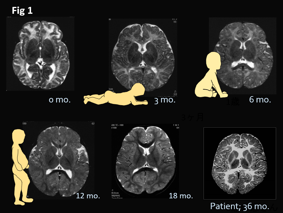
In newborn babies, white matter appears white because of its high water content (arrow). As myelination progresses, its fat content increases and gradually becomes hypointense (black) until the age of 18 months, when its appearance is almost the same as that of an adult.
White matter that appears whiter than that of a newborn is characteristic of patients with congenital cerebral hypomyelination.
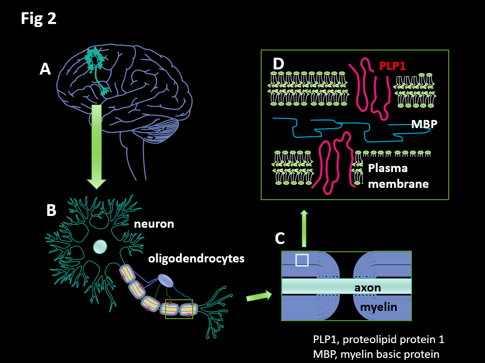
A: Conceptual diagram of a neuron in the brain. B: The axon of the neuron is surrounded by myelin (blue). C: As the axon (orange) is gradually surrounded by layers of myelin (orange) during the myelination process, it becomes capable of rapid transmission. D: The myelin wraps around the axon in layers, with adhesion factors mediating between these layers and the cell membrane; proteolipid protein 1 (PLP1), which accounts for more than half of the proteins that make up myelin, is present on the cell membrane.