Overseas Dispatch
Table of Contents
Third Retreat for Multidimensional Analysis of Memory Mechanisms at Interdisciplinary Institute for Neuroscience, Bordeaux, 2025
The Advanced Neurotechniques Course at Max Planck Florida Institute, 2025
A Graduate Student’s Long-Term Study Abroad Report, 2025
Report from Advanced Imaging Techniques for Neuroscience (AITN) @Bordeaux School for Neuronscience, 2024
Second Retreat for Multidimensional Analysis of Memory Mechanisms@Max Planck Florida Institute, 2024
Reports from Imaging Course at Max Planck Florida Institute, 2024
Third Retreat for Multidimensional Analysis of Memory Mechanisms at Interdisciplinary Institute for Neuroscience, Bordeaux, 2025
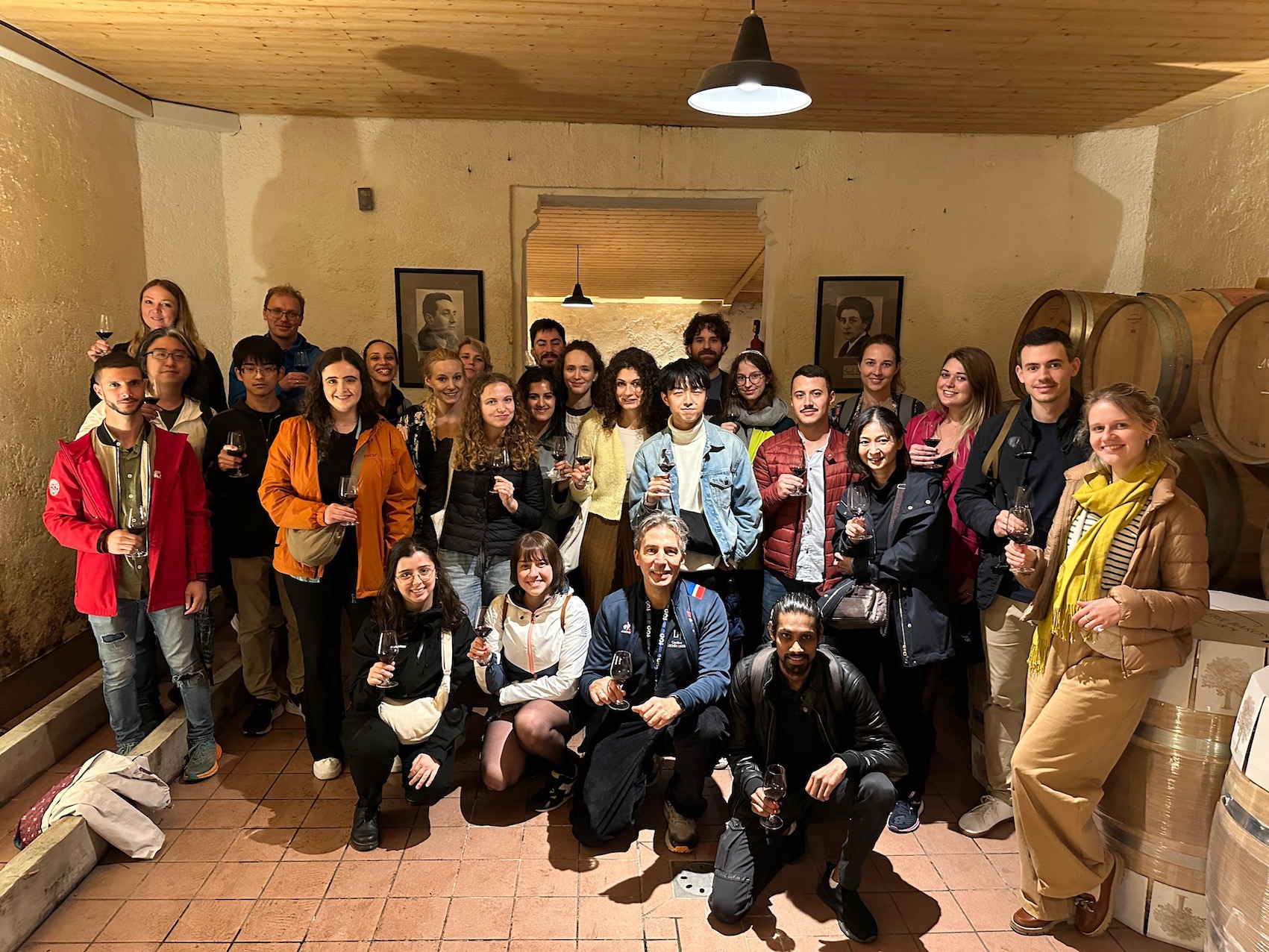
Our International Leading Research program held the third retreat at the Interdisciplinary Institute for Neuroscience, Bordeaux, Oct 20-24, 2025.
Report 1
Participant: Tomoko Fujii (Fumi Kubo Lab)
RIKEN
I had the great opportunity to participate in the “Multidimensional Analysis of Memory Mechanisms” retreat, which took place from October 20 to 24, 2025, at the IINS, Carreire Campus, University of Bordeaux.
During the symposium held on October 20–21, I was inspired by the talks given by leading neuroscientists. As I study visual processing and behavioral plasticity using zebrafish, I was particularly interested in presentations on the neural circuit mechanisms underlying plasticity in various sensory modalities, such as vision, somatosensation, and taste, mainly investigated in mice. At the same time, learning about molecular mechanisms of synaptic plasticity and the cutting-edge imaging techniques, which I do not usually encounter in my daily research, was a valuable experience. In the poster session, we had lively discussions with graduate students and postdoctoral fellows from both Bordeaux and Japan. I received insightful questions and comments on my own presentation, regardless of differences in research background or model organism, which I hope to incorporate into my future work.
From October 22 to 24, we visited the laboratories of Dr. Naoya Takahashi and Dr. Mario Carta at IINS. On the first day, we attended a joint lab meeting, where members from both groups kindly introduced their ongoing projects. We also had the opportunity to present our own research in Japan. In Dr. Takahashi’s lab, I observed behavioral experiments and neural activity imaging in the somatosensory cortex of mice. It was particularly interesting to see the experimental setup designed to measure sensory thresholds by applying subtle whisker deflections, and to observe wide-field cortical imaging using one-photon microscopy. I was impressed by their behavioral setup which is very sophisticated and systematically designed. In Dr. Carta’s lab, they showed us how acute brain slices are prepared and how patch-clamp recordings are conducted. The detailed explanations of both the experimental procedures and their scientific aims helped me better understand how the data I usually find in papers are actually obtained.
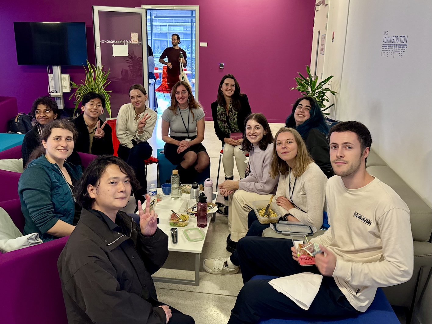
At IINS, I was impressed not only by the high level of motivation among the researchers, but also by the open and collaborative atmosphere of the institute. Many laboratories share the same floor, fostering active interaction among groups and members. I also greatly appreciated the informal conversations I had with local graduate students and postdocs, during which they shared practical tips, their daily research routines, and personal perspectives on their work.
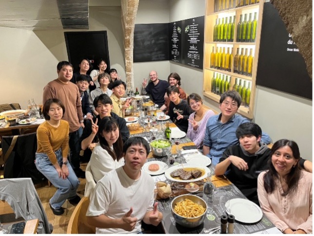
During the lab visit, Dr. Takahashi kindly organized a Q&A session on research careers, where he shared his own experiences and insights about studying abroad and career paths after finishing PhD. He was joined by Japanese postdoctoral researchers currently working at IINS, who also offered valuable advice. We continued our discussions during lunch and in the evenings, which provided an excellent opportunity to strengthen connections between researchers.
This retreat was an invaluable experience that could only be achieved through on-site participation and direct interaction. It not only broadened my knowledge and provided constructive feedback on my research, but also greatly enhanced my motivation for future studies. I would like to express my sincere gratitude to all the organizers, as well as to the faculty members and researchers at IINS, for their warm hospitality and generous support in making this visit possible.
Report 2
Participant: Shinya Oginezawa (Takayasu Mikuni Lab)
Niigata University
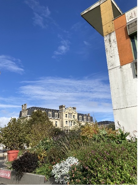
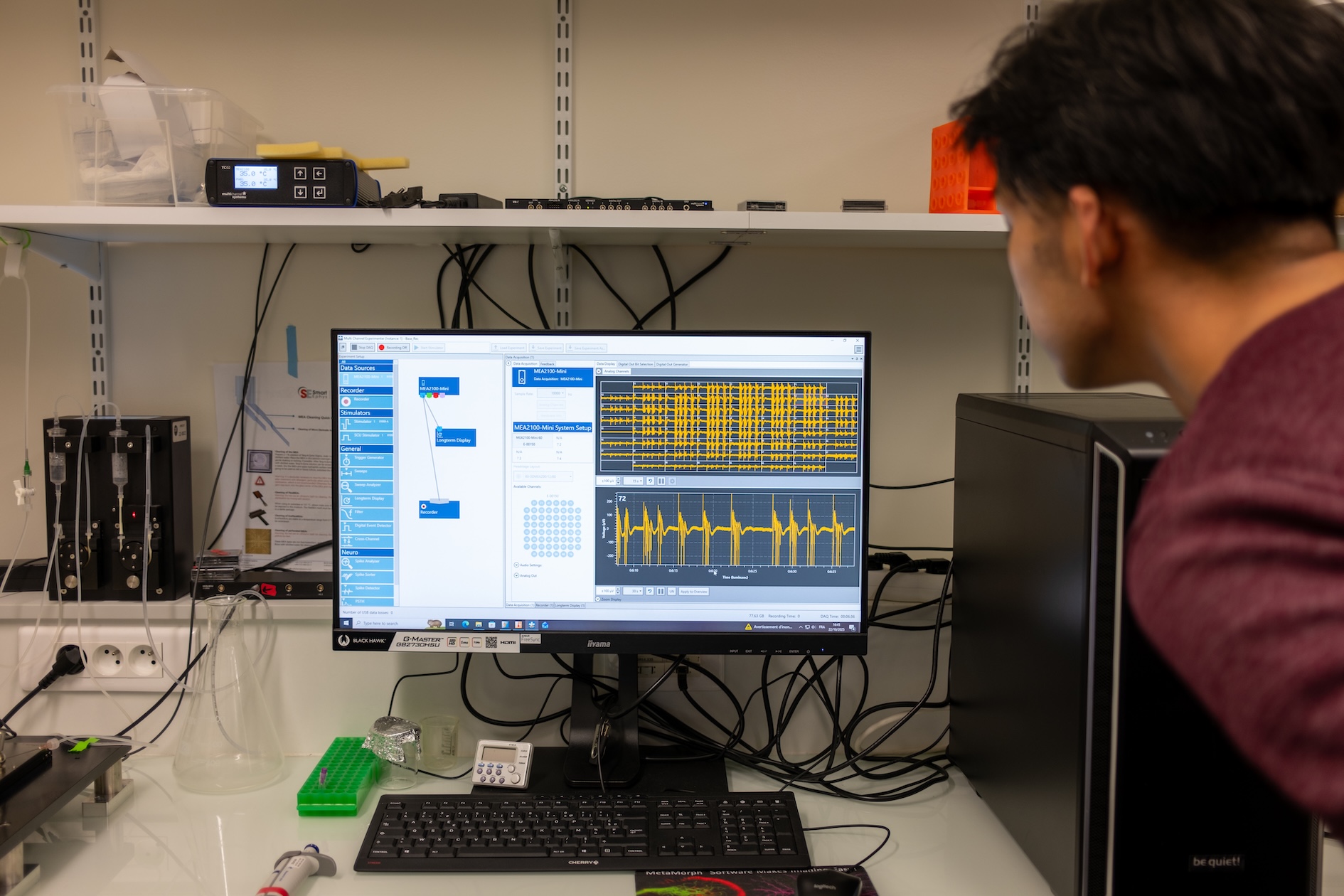
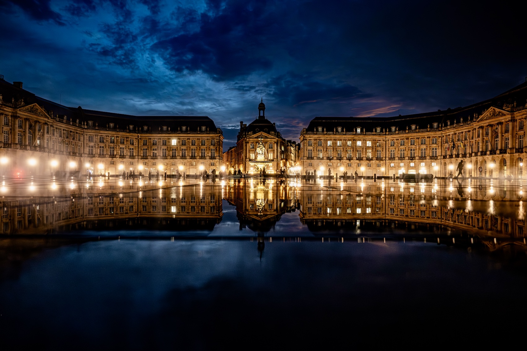
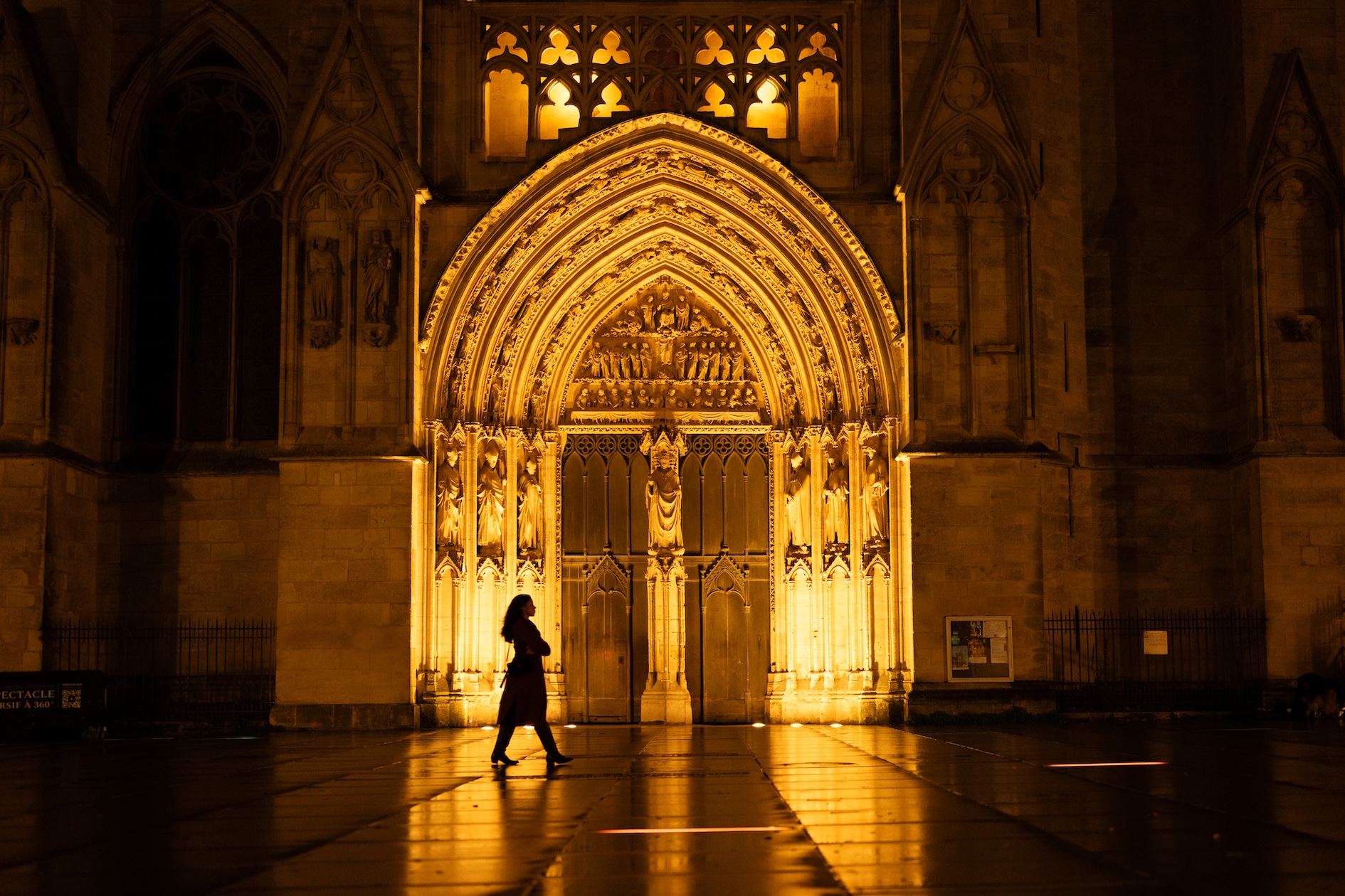
I recently had the opportunity to attend the International Leading Research Retreat held at the IINS in Bordeaux. I am now pleased to present this report detailing my experiences, what I learned, and my overall impressions. The first two days of the retreat were highly stimulating, featuring lectures by seasoned neuroscientists and research presentations by students and early-career researchers of my generation. I was able to interact closely with various researchers during coffee breaks, lunches, and especially in the evenings over a glass of Bordeaux wine. I also had the chance to present my own research via a poster. This was my first time presenting in the field of neuroscience outside of Japan, and I confess I was quite nervous. However, Professor Laurent Groc, with whom I would later spend time during my lab visit, was the very first person to come and listen to my presentation. His warm comments and strong encouragement were a huge source of motivation. The latter three days were dedicated to the lab visit. As mentioned above, I visited Professor Laurent Groc’s lab for three days. On each day, a PhD student or postdoc rotated in guiding me through the lab, giving lectures, and explaining experiments. Specifically, I was shown procedures for imaging techniques such as STORM/PALM and FRAP, as well as MEA recording. I am currently deeply interested in FRAP and had been hoping to try it myself, so the experience was extremely valuable. As the lab conducts cutting-edge research on the NMDA receptor (NMDAR), many of the experiments were related to NMDAR function, which was very informative. As a neurologist, I have treated cases of Anti-NMDAR Encephalitis, but I did not have a deep understanding of the connection between the clinical picture and the role of the NMDAR. Learning that changes in NMDAR dynamics and function affect the cognitive, memory, and psychiatric symptoms observed in the disease was truly amazing. When I shared the actual clinical picture of these patients (such as the tendency for it to affect young women and cause severe psychiatric symptoms) with the lab members, they seemed quite interested. This experience reinforced the importance of both neuroscience knowledge and clinical knowledge. Despite their busy schedules, the lab members patiently demonstrated their experiments and engaged in frequent communication during lunch and breaks, making my stay comfortable throughout. Bordeaux, where the retreat was held, is a beautiful city, and I took every opportunity to walk around with my camera, indulging in my hobby of photography. While I still feel inadequate in the research sphere, being able to converse with the people at the University of Bordeaux and in the labs through the photos I took is a fond memory. At the same time, this experience has strongly motivated me to deepen my knowledge of neuroscience and my research so that I can have more substantive discussions with them in the future. I intend to dedicate myself to further efforts. In closing, I would like to express my deepest gratitude to Professor Fumi Kubo and the other International Leading Research faculty members who planned and managed this retreat, and to my supervisor, Professor Takayasu Mikuni, who granted me this opportunity to participate. I am also wholeheartedly thankful to Professor Laurent Groc and all the members of his lab for their kind hospitality during my research visit.
Reports from Imaging Course at Max Planck Florida Institute, 2025
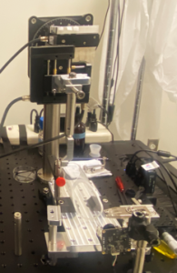
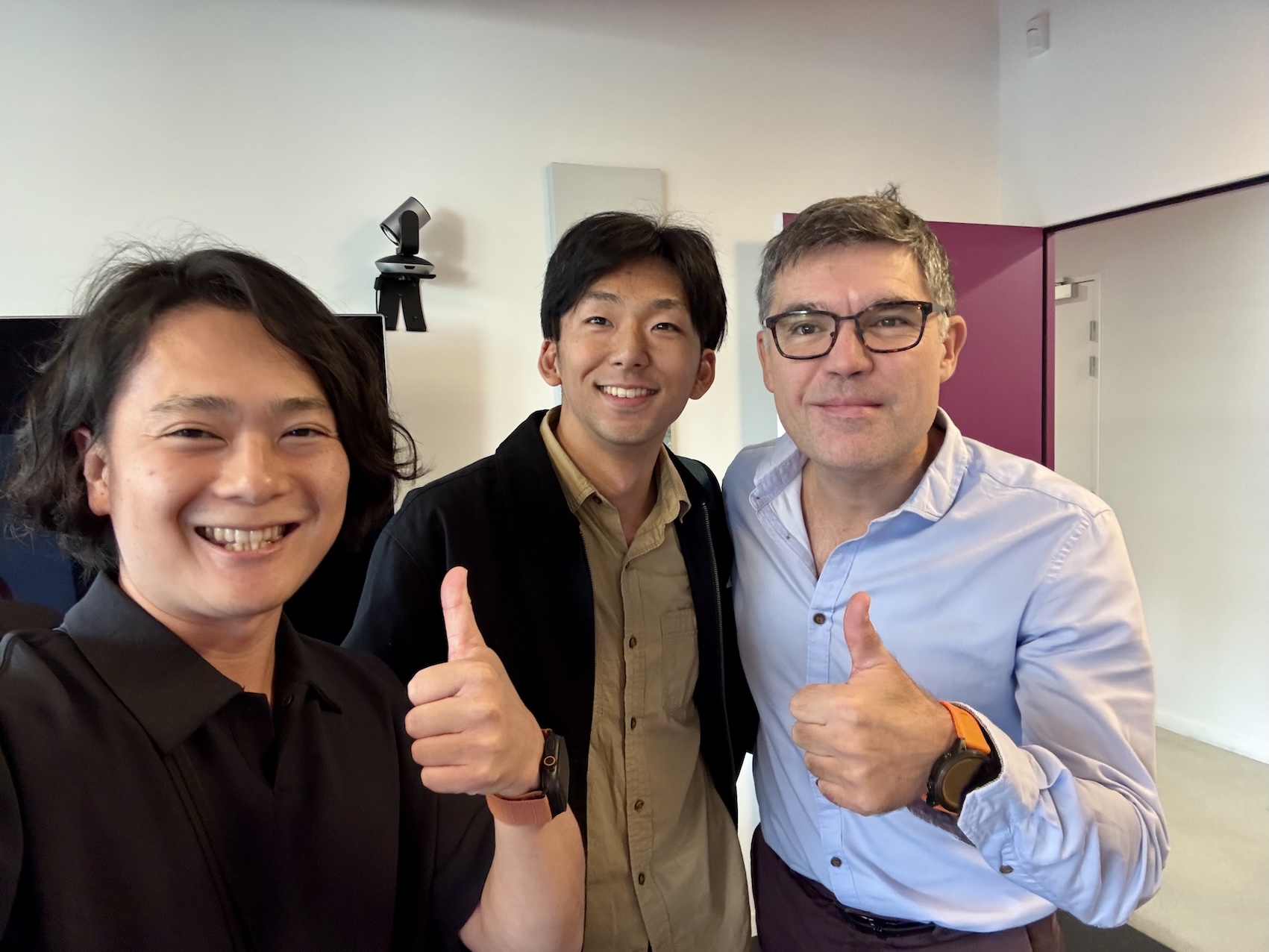
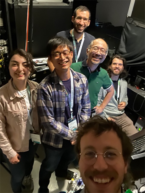
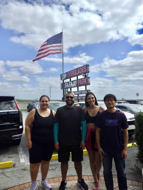
Our International Leading Research program “Multidimensional Analysis of Memory Mechanisms” supported two trainees to participate in the Imaging Course at the Max Planck Florida Institute held April 13-18, 2025.
Report 1
Participant: Ken Kunugitani (Yasunori Hayashi Lab)
Kyoto University
The Advanced Neurotechniques Course at MPFI was an ideal opportunity for me, as I am transitioning into the imaging phase of my research project. Attending this course allowed me to gain hands-on experience with cutting-edge imaging technologies and learn from world-leading experts. Through lectures and practical experiments, I learned the working principles of advanced imaging techniques and how to apply them to my own research. This knowledge will enable me to critically assess the strengths and limitations of each technology and design robust experiments tailored to my project.
The course structure consisted of morning lectures and afternoon project activities.
The morning lectures, delivered by leading experts, covered powerful techniques for visualizing and measuring the activity of neural cells and molecules, including super-resolution microscopy, biosensor for calcium and voltage imaging, holography, two-photon microscopy, electrophysiology, and FRET-FLIM.
In the afternoons, small groups conducted hands-on project activities. I chose voltage imaging/FLIM as my hands-on project. In this project, we attempted to generate a voltage-lifetime plot by correlating voltage imaging via two-photon microscopy with patch-clamp recordings, using a “chemigenetic” voltage indicator that modulates FRET efficiency through voltage-dependent changes in the absorption spectrum, developed by Dr. Ahmed Abdelfattah — a pioneering attempt worldwide. Through this project, I gained broad knowledge across multiple fields, including patch-clamp techniques, two-photon imaging, FRET-FLIM, and the use of voltage indicators. I was very glad to be part of this exciting project. I believe this experience will serve as a strong foundation as I move into the imaging phase of my research.
Beyond the technical training, I greatly enjoyed exchanging ideas about our respective research projects with other students and researchers. Learning about their diverse research approaches was highly inspiring. I also believe that the networking opportunities through this course will be instrumental as I plan to pursue postdoctoral position in the US or Europe.
Finally, I would like to express my sincere gratitude to the International Leading Research Project on Multidimensional Analysis of Memory Mechanisms for their generous support, which made my participation in this wonderful course possible. I would also like to thank my supervisor, Dr. Yasunori Hayashi for encouraging me to attend the course. Furthermore, I am deeply grateful to Dr. Ahmed Abdelfattah, Dr. Ryohei Yasuda, and Dr. Eric Salter for their invaluable guidance during the project activities.
Report 2
Participant: Sena Iijima (Yuji Ikegaya Lab)
Master’s Program, 2nd Year, University of Tokyo
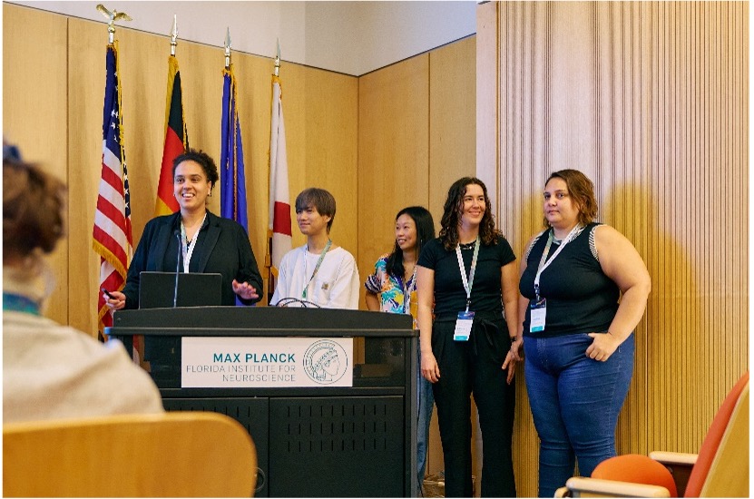
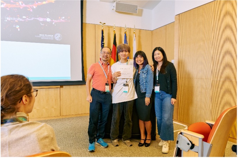
I recently had the privilege of participating in a week-long imaging course at the Max Planck Florida Institute for Neuroscience (MPFI). The course was thoughtfully designed to balance theoretical learning with practical application, featuring lectures in the mornings and hands-on project work in the afternoons.
The morning lectures far exceeded my expectations and were truly exhilarating. Leading researchers in neuroscience delivered passionate and detailed explanations of the cutting-edge technologies driving the field today. We explored everything from the principles and applications of two-photon microscopy and super-resolution microscopy to the latest biosensors, advanced electrodes, and fluorescence lifetime imaging. Hearing directly from these pioneers about the connection between theory and practice was incredibly stimulating and deeply ignited my intellectual curiosity.
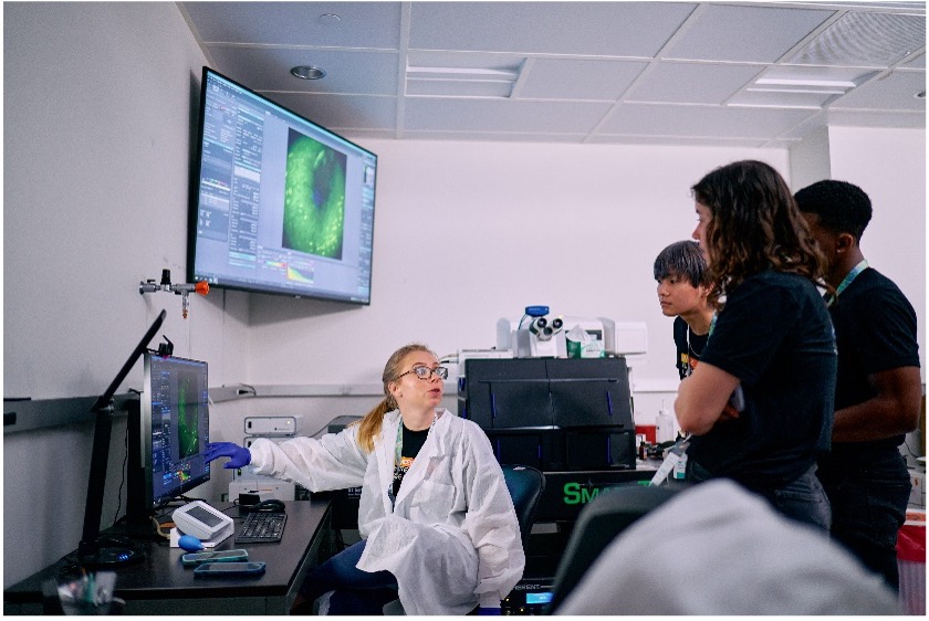
In the afternoons, we formed small groups based on our interests in project activities. I joined the “Miniscope” project, which uses a miniature microscope to observe neural activity in freely moving animals. It was an invaluable experience to work through the entire process – from assembling the Miniscope to conducting experiments with mice, acquiring data, and performing analysis – all under expert guidance. The opportunity to use state-of-the-art equipment, navigate experiments through trial and error, and ultimately present our findings as a team was profoundly impactful and a tremendous learning opportunity.
Furthermore, a remarkable aspect of this course was the high caliber of its participants. Hailing from around the world, most attendees were doctoral students or postdoctoral researchers; I was the only master’s student. The level of discussion and questioning was exceptionally high, creating an environment I found profoundly stimulating. While initially intimidated by these advanced discussions being held in English, I found all the participants to be incredibly friendly. We actively exchanged ideas about our research during breaks, lunches, and dinners – interactions the course seemed intentionally designed to foster. Encountering such diverse perspectives offered a valuable chance to re-evaluate my own research objectively while building a precious network for the future.
The wonderful Florida setting also contributed greatly to the week’s experience. Despite a demanding schedule, the excellent facilities at MPFI, combined with the beautiful natural surroundings and pleasant weather, provided the energy needed to navigate the challenging yet fulfilling days. I am confident that the knowledge, experience, and connections gained through this course will be invaluable assets in my future research endeavors.
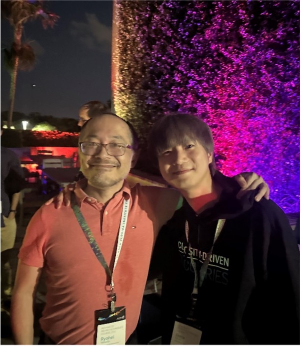
Finally, I wish to express my heartfelt gratitude to everyone at MPFI who dedicated their efforts to organizing and running this course, to Prof. Ryohei Yasuda for hosting it, and to everyone at the Cai Lab for their patient and kind guidance, especially when I faced challenges during the project. My thanks also go to everyone involved with the International Leading Research “Multidimensional Analysis of Memory Mechanisms” for their generous support, which made my participation possible. I also want to take this opportunity to extend my deep gratitude to Prof. Yuji Ikegaya for his daily guidance and warm encouragement. In return for everyone’s support, I am committed to making the most of this experience and striving to further advance my research. Thank you very much.
A Graduate Student’s Long-Term Study Abroad Report
Our International Leading Research program “Multidimensional Analysis of Memory Mechanisms” offers long-term fellowships to trainees who conduct research in labs outside of Japan.
Report
Tetsu Kawana (Yuji Ikegaya lab)
The University of Tokyo
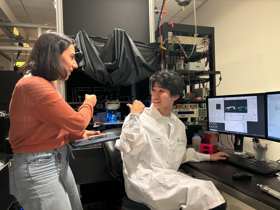
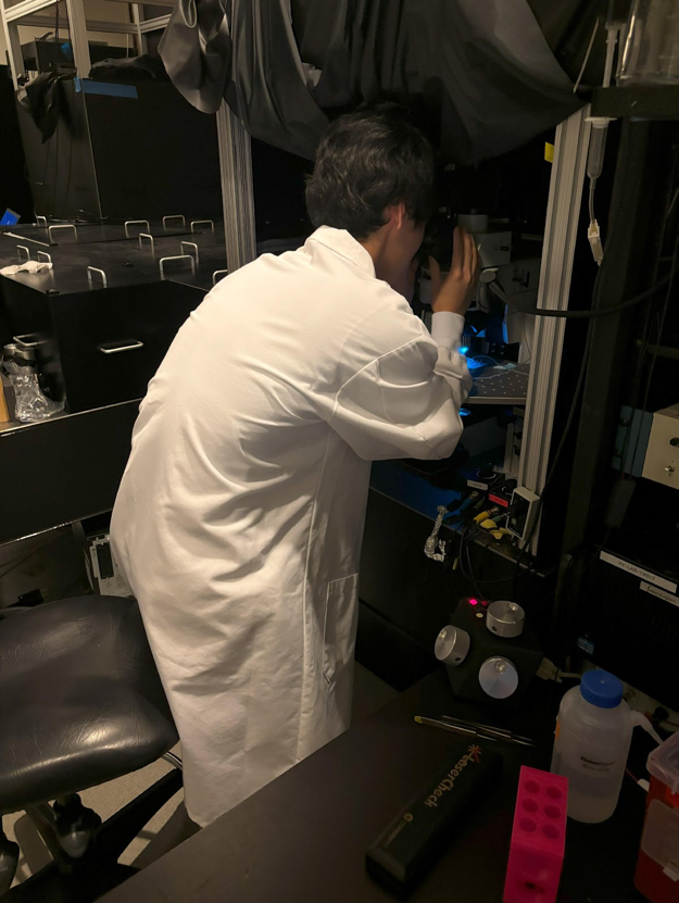
I am currently studying abroad at the Yasuda Lab in the Max Planck Florida Institute for Neuroscience (MPFI), supported by the International Leadership Research Program.
・What I am learning.
At the Yasuda Lab, I am involved in the development of a novel CaMKII biosensor and performing brain slice imaging using a two-photon microscope. I was surprised to find that nearly every researcher has access to their own two-photon microscope. Due to the customized nature of these microscopes, we must manually measure laser power and adjust the laser angles. The array of mirrors and other optical elements is remarkable. Since I have been interested in biosensor technology for a long time, I greatly enjoy working on this research. Addressing challenges such as sequence homology in fluorescent proteins within a single plasmid and finding innovative ways to improve the efficiency of FRET have made me realize the sophistication of this technology.
・What I Found Interesting About MPFI
At MPFI, external neuroscience professors and postdoctoral fellows give talks every week or two. These talks are highly valuable because they introduce diverse research topics, broadening my academic interests. After each presentation, we have a discussion over lunch with the speakers. This allows us to delve into details not covered in the talks or discuss topics such as choosing a postdoctoral fellowship (and we also get free lunch!). I make an effort to ask questions each time, and I feel that my discussion skills have significantly improved since I started.
MPFI is highly international, with researchers from various countries in Asia, such as Japan, China, Korea, and India, as well as Europe, including Italy, Turkey, Germany, and Portugal. Of course, many researchers grew up in the U.S. One of MPFI’s unique features is the opportunity to naturally form friendships with people from all over the world. Additionally, since many researchers are non-native English speakers, there is a supportive environment where people are patient with language difficulties. It is also fascinating to hear the different accents from various countries. Personally, I find Indian and Italian accents particularly challenging to understand.
・Living in Florida
Almost everything I need is available around MPFI. Occasionally, I use online shopping services like Amazon and Walmart for additional items. There is also an online supermarket called Weeee! that specializes in Asian foods, offering a variety of products, including Japanese rice, so I have no trouble preparing meals.
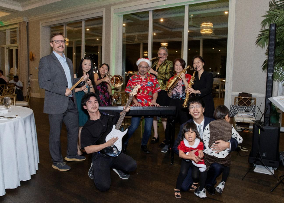
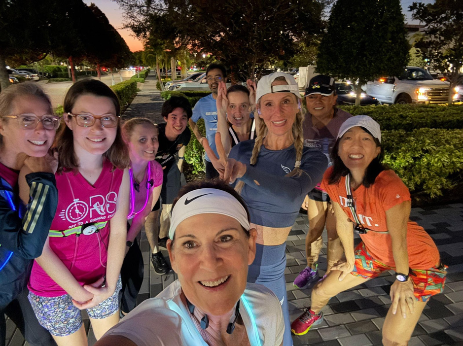
The people at MPFI are very friendly, and I quickly made friends. On weekends, I often attend parties and play board games at friends’ houses, go running, listen to music, and connect with others through shared hobbies. My research life at MPFI is incredibly stimulating, and there is far too much to describe here. Every day, I feel I am growing both personally and professionally. I remain committed to working hard to become an international researcher.
Finally, I would like to express my sincere gratitude to Dr. Ryohei Yasuda for accepting me into the lab, to the members of the Yasuda Lab for their support, and to Dr. Yasunori Hayashi, Dr. Michisuke Yuzaki, and Dr. Keisuke Yonehara of the International Leading Research for financial support. I am also deeply thankful to Dr. Yuji Ikegaya and Dr. Ryuta Koyama for permitting me to study abroad. Thanks to their support, I have gained extraordinarily valuable learning experience.

Report from Advanced Imaging Techniques for Neuroscience (AITN) @Bordeaux School for Neuronscience, 2024
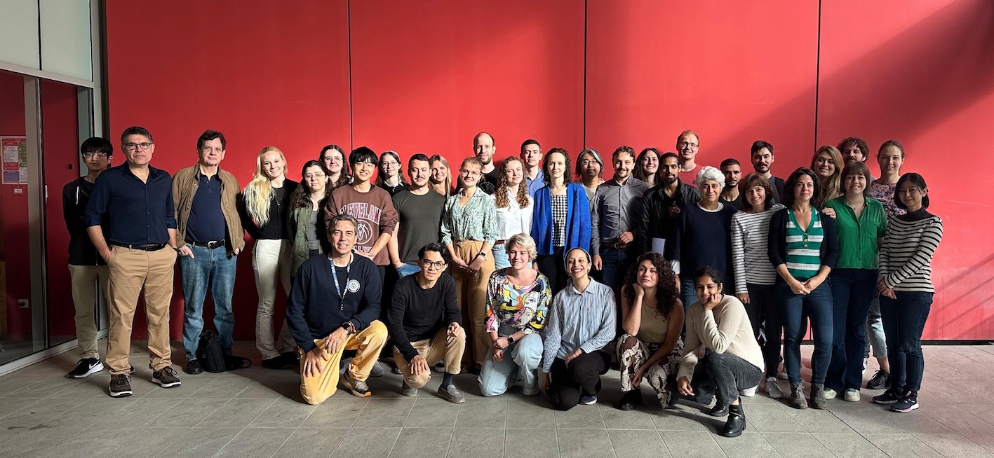
Our International Leading Research program “Multidimensional Analysis of Memory Mechanisms” supported graduate students to participate in the Advanced Imaging Techniques for Neuroscience held at the Bordeaux School for Neuronscience between September 30- October 18, 2024.
Report 1
Participant: Kazuma Miyata (Yuji Ikegaya Lab)
Master’s Program, 2nd Year, The University of Tokyo
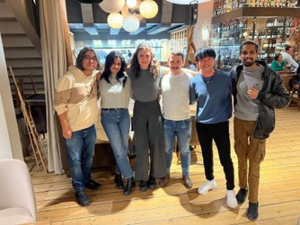
I had the opportunity to participate in the Advanced Imaging Techniques for Neuroscience (AITN) course at the Bordeaux School for Neuroscience in Bordeaux, France, as part of an international pioneering research initiative. This course focused on imaging techniques in neuroscience, both in vivo and in vitro, and offered lectures on the principles of imaging as well as hands-on training to apply these techniques over the span of three weeks. The facility was highly equipped with various imaging tools, including two-photon excitation microscopes, STED microscopes, super-resolution microscopes, and light-sheet microscopes, creating an ideal environment for learning. A total of 20 students and postdoctoral fellows, primarily from Europe, attended the course.
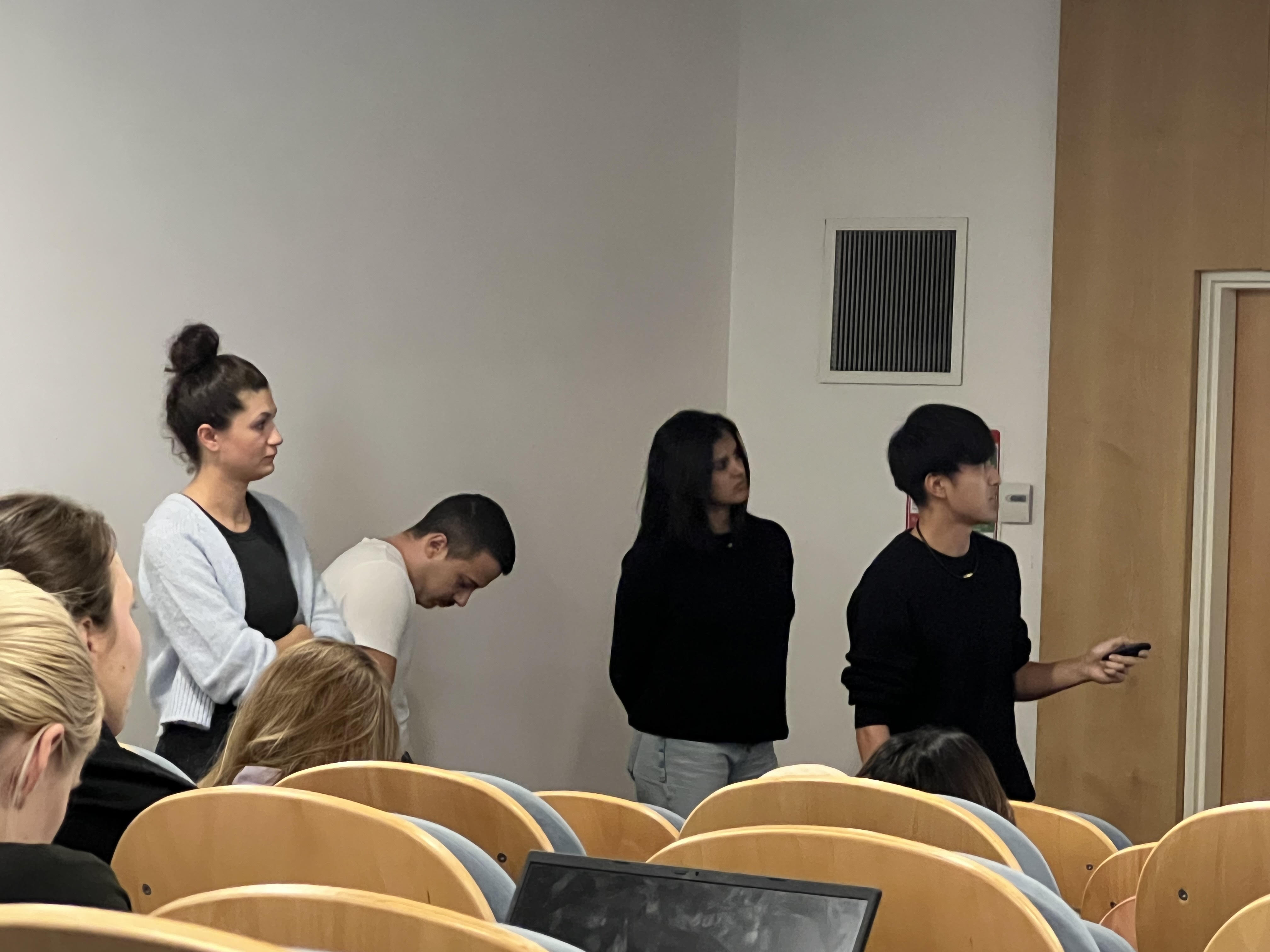
We participants engaged in separate projects during the first and second halves of the three weeks. Working with the members assigned to the same group, we conducted experiments, and at the end of each project, we had the opportunity to present our findings and knowledge gained as a group. Additionally, the course offered enriching lectures from principal investigators of research institutions from various countries, allowing us to delve deeply into the principles of cutting-edge imaging technologies.
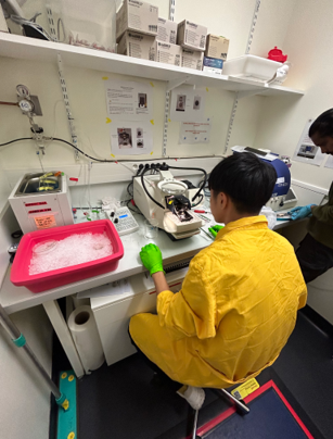
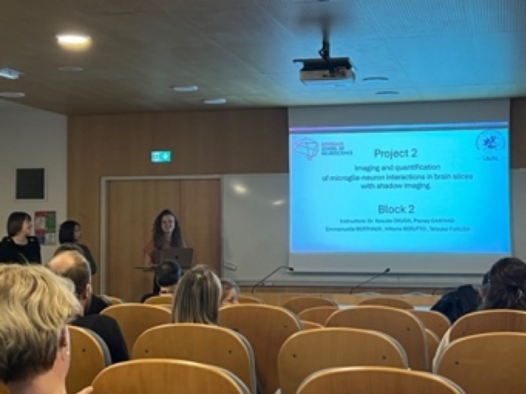
Given my interest in glial cells and Expansion Microscopy (ExM), I participated in a project on shadow imaging for microglial dynamics in the first half and on learning a new method of Expansion Microscopy in the second half. In the first project, we used mouse slice cultures and acute slices for live imaging, observing microglia extending their processes toward areas with high ATP concentrations. By combining shadow imaging—which visualizes target cells by filling the extracellular space with fluorescent dyes, rather than directly labeling the cells—with two-photon excitation microscopy, we were able to achieve highly detailed imaging. In the second project, we used a new ExM method called “LICONN,” developed by our group instructor, Dr. Mojtaba R. Tavakoli, to perform ultra-high-resolution imaging of mouse brain slices. This method involves expanding tissue twice with a water-swelling gel to increase its size by 16 times and then using deep learning-based segmentation on the tissue images to reveal complex cellular interactions. Since I use ExM in my research, I gained valuable insights on its applications and technical nuances.
Beyond the lectures and experiments, there was also time allotted for poster presentations to share our research, where I received valuable advice and had discussions with numerous researchers and students. Additionally, the course provided ample opportunities to interact with fellow participants, making it an excellent training ground for active communication in English. Through these experiences, I felt a significant boost in my motivation toward research, and I highly recommend this course to anyone interested in imaging.
Finally, I would like to express my heartfelt gratitude to Valentin Nägerl, the organizer of AITN, as well as the instructors Kosuke Okuda, Pranay Gaikwad, Nathalie Agudelo Dueñas, and Mojtaba R. Tavakoli, who taught us the experiments, Ikegaya and Koyama for their tremendous support, and the international pioneering research support team for providing me with this invaluable opportunity. Thank you very much.
Report 2
Participant: Tetsuko Fukuda (Michisuke Yuzaki Lab)
Research Associate, Keio University School of Medicine
I had the opportunity to attend the CAJAL Advanced Neuroscience Training Program on Advanced Imaging Techniques for Neuroscience, held at the Bordeaux School of Neuroscience. The course aims to promote the application and development of imaging techniques in neuroscience by exposing young researchers to a wide range of imaging concepts and practical techniques through the participation of PIs and researchers from countries that are leading the development of cutting-edge imaging techniques in neuroscience research.
The program included both imaging lectures and hands-on training in experimental techniques and data analysis, with a jam-packed three-week schedule. Lectures by developers of state-of-the-art imaging systems and leading researchers in the field helped us understand the basic principles behind different types of microscopes, the historical development of these technologies, and the latest advances, including their results. I found it particularly fascinating to learn about the development of techniques I had not previously encountered, such as light-sheet microscopy. During the practical training, there were 10 different projects, and students could choose one project for the first half and another for the second half of the course. Each project was conducted in small groups of 2–4 students, with dedicated instructors providing detailed guidance at every step.
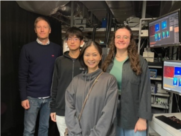
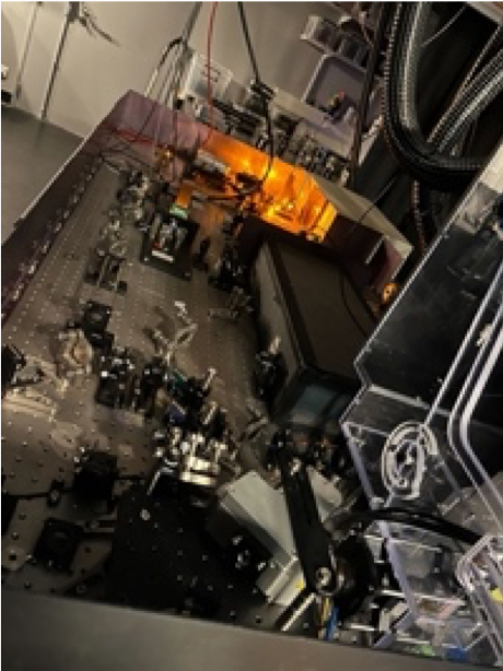
My main goal was to learn about the structure and imaging methods of STED microscopy, especially shadow imaging in vitro. Therefore, in the first half of the course I chose the SUSHI live imaging project using slice cultures, and in the second half I participated in the shadow imaging project using acute and cultured brain slice preparations, and fixed sections with two-photon microscopy. The STED microscope we used in the first half was a homemade system, and I greatly benefited from the opportunity to see its optical design up close, while receiving clear explanations of the various modifications made during its construction. I also had the opportunity to gain hands-on experience building the optical path for the laser system and aligning the laser lines, which was both fun and educational. The small class size allowed ample time for student and faculty interactions, not only to discuss the course projects, but also to exchange ideas about the research being conducted in our respective labs. I received invaluable advice from the instructors on analyzing imaging data from my own projects, as well as guidance on solving issues related to custom microscope components. I feel that participating in this course, which allowed me to learn broadly and deeply about different types of imaging and broaden the scope of my own research, was an incredibly meaningful experience.
Finally, I am deeply grateful to Professor Valentin Nägerl, the director of this wonderful course, for his many insights and advice on my research; to Professor Jan Tønnesen, Professor Kosuke Okuda, and Professor Pranay Gaikwad for their detailed guidance and advice on experiments and analysis; and to all the staff for their support. I would also like to express my sincere gratitude to Professor Yuzaki for providing me with this invaluable opportunity, to the members of my research laboratory for their support, and to the International Leading Research Project on Multidimensional Analysis of Memory Mechanisms for tremendous support.
Report 3
Participant: Ikuma Nakagawa (Keisuke Yonehara Lab)
First-year Ph.D. candidate (five-year program), SOKENDAI/National Institute of Genetics
I got a fantastic opportunity to participate in the “CAJAL course on Advanced Imaging Techniques for Neuroscience 2024”, held at the Bordeaux School of Neuroscience in Bordeaux, France, from September 30 to October 18. The course aimed to teach students the principles of cutting-edge imaging techniques and provide them with hands-on training through experimental projects to acquire technical skills. In the mornings, we attended lectures by keynote speakers and instructors, learning about the fundamentals of imaging techniques and their application in various research fields. In the afternoons and evenings, we conducted experiments in small groups of 2 to 4 students.
The three-week course was divided into two parts, with each part focusing on different experimental projects to learn the latest imaging techniques. The imaging techniques covered in the course ranged from two-photon excitation microscopy, stimulated emission depletion (STED) microscopy, light-sheet microscopy, shadow imaging, to expansion microscopy. At the end of each experimental project, each group gave a presentation to share what we had learned.
During the first half of the course, I learned super-resolution shadow imaging (SUSHI) using STED microscopy. Under the guidance of Dr. Jan Tønnesen, who developed SUSHI in 2018, we performed experiments on mouse hippocampal organotypic slice cultures. Shadow imaging involves staining the extracellular region with a fluorescent dye called Calcein, allowing us to visualize the cells as shadows and obtain information about both the cells and the extracellular space simultaneously. By adding Calcein directly to the buffer in which the sample was immersed and waiting for a few minutes, we could visualize the extracellular space and perform shadow imaging. We were able to observe spines, synapses, and the expansion of the extracellular space when osmotic challenge is applied.
In the second half of the course, I learned expansion microscopy. This technique involves embedding the sample in a gel that absorbs water and expands, chemically expanding both the gel and the sample itself. This allows for super-resolution imaging using confocal microscopy. Under the guidance of Dr. Mojtaba Tavakoli and Dr. Nathalie Agudelo Duenas, who had recently completed their PhDs, we expanded mouse brain slice samples using two different protocols and observed the cells with confocal microscopy. First, we used the traditional Protein Retention Expansion Microscopy (ProExM) method to observe neurons expanded about four times. Then, we used a new protocol developed by Dr. Tavakoli and colleagues, called LICONN (light-microscopy based connectomics), which utilizes expansion microscopy to reveal neural connections at the single-synapse level. By performing pan-protein staining and expanding the samples twice, we were able to observe intracellular structures at a 16-fold expansion.
This training course provided me with valuable hands-on experience in super-resolution microscopy techniques, which I had been particularly interested in. It greatly increased my motivation for my own research. I plan to incorporate the experimental methods I learned in this course into my own research and advance it further. Additionally, since this was my first overseas experience, the three weeks spent conducting experiments at a research institute in Bordeaux, interacting and discussing in English with instructors and other participants, was an invaluable experience for me. I hope to use this experience to make my future research during my PhD even more productive.
Lastly, I would like to express my deep gratitude to everyone at the Bordeaux School of Neuroscience for their support of the training course, to the instructors Dr. Jan Tønnesen, Dr. Mojtaba Tavakoli, and Dr. Nathalie Agudelo Duenas, to Dr. Keisuke Yahara for recommending and supporting my participation, and to the support from the International Leading Research program. Thank you very much.
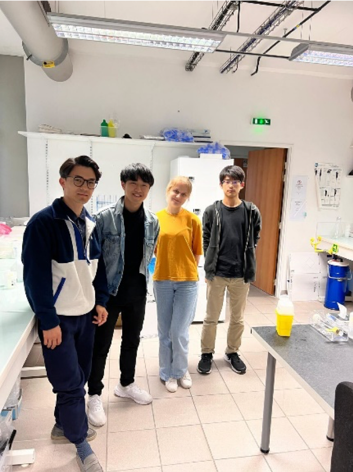
Second Retreat for Multidimensional Analysis of Memory Mechanisms
We held the Second Retreat for Multidimensional Analysis of Memory Mechanisms at the Max Planck Florida Institute between September 18-21, 2024. Students and postdocs stayed for 3 more days for lab visits.
Report 1
Participant: Joe Sakamoto (Tomomi Nemoto Lab)
Project Research Staff, National Institute for Physiological Sciences
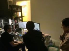
I had the opportunity to participate in the Memory Formation Retreat at the Max Planck Florida Institute for Neuroscience (MPFI) followed by a lab visit. During the retreat, the world’s leading neuroscientists, graduate students, and early career researchers gave great presentations. I also gave an oral presentation in the Young Session and presented a poster. After my talk, I received valuable feedback from the attendees. I’d like to incorporate the feedback from the discussions with the attendees to further improve my research. The retreat included a tour of MPFI’s machine shop and Imagine Center, and I could see many state-of-the-art equipment in the tour. In the social activity session, I visited the Loggerhead Marine Life Center, a sea turtle care center, and Juno Beach with several retreat participants and made good connections.
On September 22, Dr. Ryohei Yasuda held a career seminar. He invited Dr. Lin Tian and Dr. Hidehiko Inagaki to discuss career development in the United States, including opportunities for applying for research grants after obtaining a Ph.D. degree. After the seminar, we seamlessly started a social hour with the seminar participants and discussed on a wide range of topics, such as our research, hobbies, astronomy, physics, and so on.
Starting on September 23, I visited Dr. Ryohei Yasuda’s lab for three days with two other visitors from Japan. In the morning of the first day, we attended a lab meeting and a journal club, followed by a lecture on the basics of optical and two-photon microscopy in the afternoon. Since I give similar lectures about microscope in Japan, the lecture was very helpful for me. From the 2nd day, Dr. Tristano Pancani, Irena Suponitsky-Kroyter, Goksu Oz in Dr. Yasuda’s lab, Koichi Hirokawa in Dr. Inagaki’s lab showed us several experiments, such as acute brain slice and organotypic slice preparations, LTP induction using two-photon uncaging with caged glutamate, accompanying electrophysiology and fluorescence lifetime imaging experiments, and behavioral experiments. Dr. Yasuda’s lab has unique and sophisticated microscope setups, which is why I chose Yasuda’s lab for my lab visit. It was a great opportunity to ask and discuss some specific questions in front of their microscopes. Especially, the measurement of physiological function using 2P-FLIM with various probes is a unique technique in Yasuda’s lab, and learning about their setup in detail was a significant gain for me.
Finally, I would like to express my gratitude to Dr. Takayasu Mikuni, Dr. Ryohei Yasuda, and all the staff members who organized and conducted this retreat, and to Dr. Tomomi Nemoto for giving me the opportunity to stay at MPFI. I also would like to thank people who showed us their experiments, Dr. Yoshihisa Nakahata for organizing our lab visit program, and Dr. Ryohei Yasuda again for allowing us to visit his laboratory. It was a great experience for me to learn about the many latest research, advanced laboratory facilities, and experimental techniques at MPFI by participating in this retreat. I believe that the experience of this retreat and lab visit can enrich my own research life.
Report 2
Participant: Shambhavi Chaturvedi (Keisuke Yonehara Lab)
Third-year Ph.D. candidate (five-year program), SOKENDAI/National Institute of Genetics
I had the privilege of attending the Multidimensional Memory Mechanisms Retreat, organized by the Max Planck Florida Institute (MPFI) in Jupiter, Florida. The retreat was divided into two key segments: a scientific conference and lab visits for students.
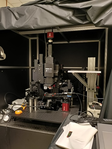
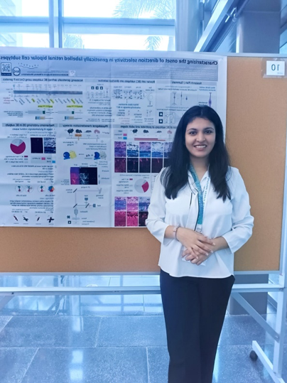
During the two-day conference, I attended a series of intriguing presentations that spanned a broad spectrum of topics, ranging from advances in molecular genetics to insights into complex neural networks and disease mechanisms. These sessions were further enriched through engaging discussions ranging from hot topics in current neuroscience research to broader themes, such as the study of consciousness with highly accomplished supervisors during breaks. We also had the opportunity to visit MPFI’s impressive machine room, which is well-equipped to build various instruments for research. The retreat also included poster sessions, which helped to exchange ideas and gain fresh perspectives on several ongoing projects. I had the opportunity to learn about advancements in two-photon microscopy, emerging imaging technologies, novel sensor engineering, and experimental approaches in physiology and behavior.
Following the conference, I visited Dr. Lin Tian’s lab to explore the latest developments in sensor technology and neural therapeutics. The lab visit was structured across three days:
- Day 1 focused on an introduction to light-sheet microscopy and in vitro experiments. I also attended a lab meeting where the use of dLight sensors in non-traditional brain regions, such as the medial prefrontal cortex (mPFC) and nucleus accumbens (NAc), was discussed. I observed experiments in which the dopamine sensor injected into the NAc was monitored using a 1-photon miniscope setup.
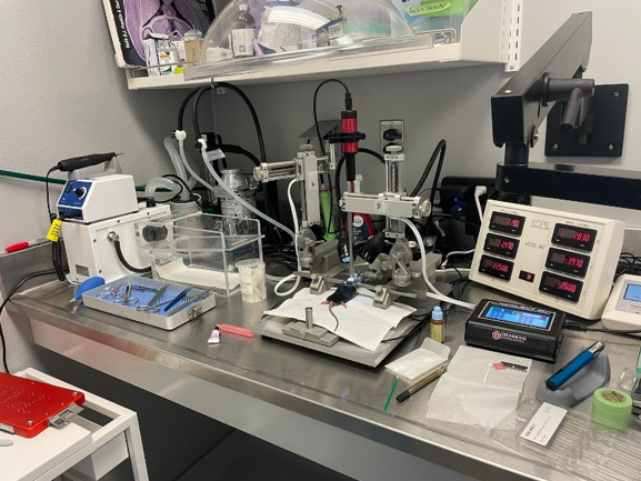
- Day 2 started with an introduction of a current lab project, which focuses on developing a splitable fluorescent iGluSnFR sensor designed to capture circuit-specific glutamate transients at synapses. I also observed surgical procedures related to the delivery of a serotonin sensor (iSeroSnFR) in orbitofrontal cortex.
- Day 3 involved practical demonstrations of vibratome sectioning to prepare striatal samples and two-photon imaging to visualize signals from injected sensors in striatal neurons.
This lab visit enriched my understanding of various novel sensors, 2 photon imaging, and behavioral neuroscience techniques. In addition, insightful discussions with lab members deepened my knowledge of the applications of these tools in future research.
I am very grateful to Dr. Lin Tian for providing the opportunity to engage with the innovative research conducted at her lab. I am also grateful to Akash Pal, Yihan Jin, Kiran Long-Iyer, Julie Chouinard, and all the members of the Tian Lab for demonstrating their experiments and offering guidance and support throughout my visit. Overall, the retreat was an enriching experience, significantly broadening my scientific knowledge and allowing me to connect with many people in the field. I am especially grateful for the guidance I received on academic career journey from Dr. Ryohei Yasuda, Dr. Hidehiko Inagaki, and Dr. Lin Tian. Lastly, I would like to express my sincere gratitude to my supervisor, Dr. Keisuke Yonehara, for encouraging me to attend the conference. I am also very thankful for the generous funding provided by the International Leading Research Project on Multidimensional Analysis of Memory Mechanisms, which enabled my participation in this memorable retreat. I look forward to applying the insights gained from this experience to my own research.
Reports from Imaging Course at Max Planck Florida Institute, 2024
Our International Leading Research program “Multidimensional Analysis of Memory Mechanisms” supported two graduate students to participate in the Imaging Course at the Max Planck Florida Institute held February 5- February 17, 2024.
Report 1
Participant: Chung-Han Wang (Fumi Kubo Lab)
Third-year Ph.D. candidate (five-year program), SOKENDAI/RIKEN
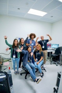
I had the opportunity to join the Neuroimaging course held by Max Planck Florida Institute for Neuroscience (MPFI). The MPFI campus, located in Jupiter, Florida, is surrounded by the Wertheim UF Scripps Institute and Florida Atlantic University (FAU). The MPFI neuroimaging course was an intensive and comprehensive laboratory-oriented course focusing on applying imaging techniques for neuroscience research. The course was structured to include morning lectures and afternoon/evening laboratory sections, featuring lectures by MPFI PIs and invited professors. This time, there were a total of 22 course participants, including PhD students and postdocs.
The first afternoon featured an ice-breaking activity to help everyone get acquainted. The second day was particularly significant, as all students were divided into four teams for station rotations (lab rotations). There were ten stations to choose from, including mini-2photon microscopy, holographic optogenetic stimulation, 3-photon microscopy, voltage imaging, miniature microscopy in moving mice, DNA PAINT, mini-2photon in FLIM/FRE, and electron microscopy (EM). The morning sessions began with lectures on optics, microscopy basics, advanced microscopy techniques, and imaging, aiming to provide students with a comprehensive understanding—from fundamental principles to practical applications like laser alignment. All lectures were delivered by established researchers in their respective fields, including several invited PIs who were the inventors of topics such as mini-2P and Voltron. Additionally, on two evenings during the course, we held data blitz sessions where students presented their PhD projects in five minutes.
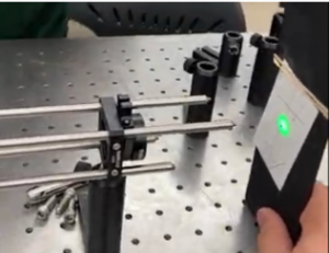
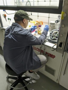
In my case, I was allocated to the EM station with another student due to our shared interest in relevant research. Since the participants assigned to the EM station were predetermined, Dr. Naomi Kamasawa, Head of the MPFI Imaging Center, inquired if we wished to bring our own samples. This allowed me to send my fixed zebrafish from Japan to MPFI for serial block-face SEM. At the EM core, we learned about various aspects of sample preparation for EM, including enhancing sample contrast, heavy metal staining, dehydration for resin infiltration and ultrastructure preservation, microtome shaving and sectioning, trimming the resin sample to fit with the pin, and coating to prevent charging—all under the guidance of research scientist Debbie Guerrero-Given. We gained a deeper understanding of the structural and functional differences between SEM and TEM and had the opportunity to capture images using both techniques.
I highly recommend people to consider applying for this imaging course. The imaging course offers an exceptional opportunity for individuals (not only students but PIs also) who are eager to explore the latest technologies and methodologies in neuroimaging. Lastly, I am deeply grateful to my supervisor Dr. Fumi Kubo and Dr. Keisuke Yonehara for the encouragement to participate in the course. Furthermore, I extended my sincere appreciation for the generous financial support from the International Leading Research Project on Multidimensional Analysis of Memory Mechanisms, which made my participation in this unforgettable course possible. Additionally, I would like to express my thanks to Naomi and Debbie for their invaluable assistance and guidance during the MPFI two-week course, which significantly contributed to my learning and the enrichment of my understanding in EM research.
Report 2
Participant: Shiyori Tanaka (Yuji Ikegaya Lab)
Second-year Ph.D. candidate, University of Tokyo
I had the opportunity to participate in the Imaging Course held at the Max Planck Florida Institute (MPFI) for two weeks. This course is designed with the aim of incorporating imaging techniques into one’s own research and advancing it further. The participants ranged from undergraduate students to PhD candidates and postdocs, creating a highly diverse environment where people from all over the world could learn together.
The Imaging Course consisted of morning lectures and afternoon lab sessions, with the lectures covering a wide range of topics related to imaging. The curriculum began with the basic principles of optics, and from this foundation, we systematically learned advanced imaging techniques, which made the content highly understandable and applicable to a wide range of areas. What left the greatest impression on me was how the curriculum seamlessly connected theoretical knowledge with practical experiments, effectively bridging theory and practice. This allowed me to approach the experiments with a solid understanding of the technical background.
During the lab sessions, participants were divided into small groups of two to three and assigned to different research labs. I was fortunate to be selected for my first-choice project, “Mini2P,” where I worked on a project to observe neural activity in freely moving mice using a Mini2P, a small two-photon microscope. This project was hosted by the Ryohei Yasuda Lab and Yingxue Wang Lab at MPFI. We were guided by Weijian Zong, the developer of Mini2P, as well as by doctoral students Goksu Oz, Zhuoyang Ye, and Ohm Parikh. Under their supervision, I learned the entire process, from setting up the Mini2P to recording neural activity.
In addition to research, I was able to enjoy the natural beauty of Florida during the course. The environment around MPFI is excellent, with vast grounds and clear air. During breaks from research, I took the opportunity to relax by visiting the beach, exploring local bars, and enjoying street performances. The hotel where I stayed was also very comfortable, which helped me maintain my health and focus, despite the intense schedule that ran from 9 AM to 9 PM every day.
Through this course, I was able to engage with cutting-edge imaging technologies and expand my network with researchers around the world. I joined this course motivated by my desire to incorporate imaging technology into my current research theme and to observe neural activity in detail during patterned light stimulation. Thanks to the knowledge and skills I gained from this course, as well as the connections made with colleagues from around the globe, I feel that the path to achieving my goals has become clear.
Finally, I would like to express my deep gratitude to everyone at MPFI who provided me with this invaluable experience, especially Dr. Weijian Zong, Dr. Ryohei Yasuda, Dr. Yingxue Wang, the instructors, TAs, and Julia Dziubek, who supported and encouraged me throughout the course, as well as the professors and seniors in Japan who have supported me, especially Dr. Yuji Ikegaya. I will continue to advance my research, drawing on the experiences gained from this course. Thank you very much for your support.
