|
NEJM勉強会2003 第11回03/04/16 実施 Aプリント 担当:宮垣朝光 Case 4-2003: A 42-Year-Old Woman with Cough Fever and Abnormalities on Thoracoabdominal Computed Tomography (Volume 348: 447-455)
【主訴】 湿性咳漱、寒気、発熱 【現病歴】 入院の7ヶ月前までは健康であった。その頃より断続的な乾性咳漱が出現し、抗生物質投与にもかかわらず改善しなかった。しかし、CXpでは異常所見は見られなかった。2,3ヶ月前再度CXpを施行し、裂孔ヘルニアと診断されたが造影では指摘できなかった。2,3日前より緑色の悪臭を伴う痰を喀出し初め、38.5℃の発熱が見られた。但し、寒気、汗は見られなかった。CXpでは右下葉に空気〜液体レベルの円形領域が見られた。そして、入院となった。 【生活歴】 生粋のアルバニア人で都市部に居住していた。会計士をしており、農家(ほとんどは羊を飼っていた)との取引のため、毎日農村部に出かけていた。アルバニアを離れる1年前(入院の3年前)に都市部生まれの犬を飼い始めた。犬との接触はそれ以前はなかった。入院の2年前に家族でアメリカ合衆国に移住し、それ以降は事務職についていた。移住前のツ反(-)。アルコール(-)、smoking(-)、常用薬剤(-)、海外旅行歴(-)。 【既往歴】 アルバニアにて長年反復性の気管支炎に罹患していた。他に特記すべきことなし。 【家族歴】 特記すべきことなし。 【現症】Admission on foot <VITAL SIGNS> BT 37.2℃, BP 100 / 60 mmHg, PR 118 /min, RR 20 /min, SaO2 97 % (room air) <HEAD> [Eyes] 左目の硝子体が先天的に不透明. Retina:n.p. [Oral cavity] tongue:n.p., 歯科衛生は悪く、齲触、欠けた歯が見られた. [Neck] no goiter, cervical bruit(-) <LUNG> 打診にて右肺底部に濁音あり., normal vesicular sound, no crackles, lung sound clear <HEART>Ⅰ→Ⅱ→Ⅲ(-),Ⅳ(-), LevineⅠ/Ⅵ systolic murmur <ABDOMEN> n.p. <LYMPHATICS> neck / supraclavicular / axillary / inguinal:not swollen <EXTREMITIES> n.p. <NEUROLOGICAL> n.p. 【入院時検査所見】 <CBC> Neu 68%, Eos 5%, Baso 1%, Mono 14%, Lym 12%, Ht 30.9%, MCV 76 fl, Plt 27.9×10⁴ /μl <CHEMISTRY> BUN normal, Cr normal, Na 144 mEq/l, K 4.3 mEq/l, Cl 108 mEq/l, HCO3- 28 mEq/l, FBS 113 mg/dl, Fe 12 μg/dl, TIBC 320 μg/dl, Transferrin 256 mg/dl, Ferritin 46.2 ng/ml <ECG> HR 104 /min, sinus tachycardia, 小さな非特異的ST segment, abnormal T <CT> 右下葉の背底区に空気〜液体レベルの6x5cm程度の厚い壁を有した嚢胞性の構造が見られる(Fig.1)。肝右葉に13x11x9cm程度の境界明瞭でlow densityなmassを認める。嚢胞性で多房性。細長い筋が散見される。また、大きな嚢胞の中に境界明瞭な小さい嚢胞が見られる(Fig.2)。その他の胸腹部臓器は正常。 診断と手技をお願いします。 
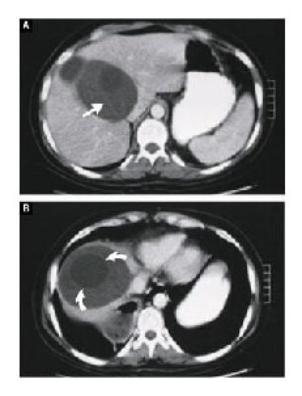
Figure 1. Contrast-Enhanced Thoracic CT Scans. (上図左) Panel A is a lung window, and Panel B a soft-tissue window. In the right lower lobe, there is a thick-walled structure with an air?fluid level. The dome of the diaphragm is seen above the cavity. Figure 2. Contrast-Enhanced Abdominal CT Scans. (上図右) A large cystic lesion containing streaks of debris (arrow) is present in the right lobe of the liver (Panel A). A view at a lower level shows what appears to be a cyst within a cyst (curved arrows) (Panel B). |